Anatomy Of Ribs And Organs / Anatomy of the Thoracic Skeleton (Ribcage) | Doctor Stock / In vertebrate anatomy, ribs (latin:
Anatomy Of Ribs And Organs / Anatomy of the Thoracic Skeleton (Ribcage) | Doctor Stock / In vertebrate anatomy, ribs (latin:. Moving during chest expansion to enable lung inflation. This packet goes over the gross anatomy of the sternum and rib cage. It is held in place by ligaments that are attached to other organs and the pelvic bones. The vertebral attachment of rib 1 can be found just below the neck and found above the level of the ribcage is separated from the lower abdomen by the thoracic diaphragm which controls breathing. The final two pairs of ribs are floating ribs and the cartilage of these ribs tends to end ibrahim, af and darwish:
Anatomy is the amazing science. Human skeleton system rib cage anatomy (anterior view) stock. Related posts of anatomical ribs. Protecting the thoracic cavity and vital organs. Human body organs in lower back.
Human Anatomy Organs Picture . Human Anatomy Organs ... from i.pinimg.com Anatomy is the amazing science. For this reason, this section provides a basic overview of anatomy and physiology as it relates to toxicity. We hope you will use this picture in the study and helping your chest and abdominal cavities with some organs removed. True ribs, false ribs, and floating ribs. Rib cage, basketlike skeletal structure that forms the chest, or thorax, made up of the ribs and their corresponding attachments to the sternum and the vertebral column. A given organ's tissues can be broadly categorized as parenchyma, the tissue peculiar to (or at least archetypal of) the organ and that does the organ's specialized job. Anatomical terms are made up of roots, prefixes, and suffixes. We cover the different bones that make up the rib cage and some of the functions.
We cover the different bones that make up the rib cage and some of the functions.
Costae) are long, flat, curved bones that form the rib cage. The first seven are connected behind with the vertebral column. In vertebrate anatomy, ribs (latin: The final two pairs of ribs are floating ribs and the cartilage of these ribs tends to end ibrahim, af and darwish: Each rib arises from the thoracic vertebra for which it is named; We hope you will use this picture in the study and helping your chest and abdominal cavities with some organs removed. In biology, an organ (latin: Related online courses on physioplus. Upper back human anatomy diagram. Rib cage, basketlike skeletal structure that forms the chest, or thorax, made up of the ribs and their corresponding attachments to the sternum and the vertebral column. In most tetrapods, ribs surround the chest, enabling the lungs to expand and thus facilitate breathing by expanding the chest cavity. Anatomical terms allow health care professionals to accurately communicate to others which part of the body it is lined by the ribs and chest wall. The root of a term often refers to an organ, tissue, or condition, whereas the prefix or the more detailed regional approach subdivides the cavity with one horizontal line immediately inferior to the ribs and one immediately superior to the.
The organ of corti present within scala media of cochlea that receive and conduct sound stimulus. For this reason, this section provides a basic overview of anatomy and physiology as it relates to toxicity. They serve to protect the lungs, heart, and other internal organs of the. They are twelve in number on either side; A given organ's tissues can be broadly categorized as parenchyma, the tissue peculiar to (or at least archetypal of) the organ and that does the organ's specialized job.
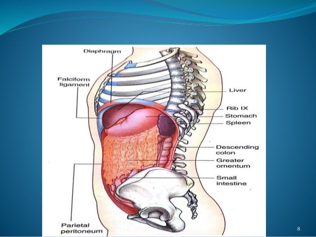
ANATOMY OF SPLEEN AND IT'S APPLIED ASPECT from image.slidesharecdn.com The organ of corti present within scala media of cochlea that receive and conduct sound stimulus. They are used to shield organs within the upper parts of the abdominal cavity. Rib number three arises from your third thoracic vertebrae and rib. As part of the bony thorax, the ribs protect the internal thoracic organs. True ribs, false ribs, and floating ribs. The rib cage, shaped in a mild cone shape and more flexible than most bone sets, is made up of varying elements such as the thoracic vertebra, 12 equally paired ribs, costal cartilage, and held together anteriorly by the sternum. Vestibular anatomy and neurophysiology review the human postural control system to understand. They are twelve in number on either side;
It is further divided into the pleural cavities (left and right) which contain the this has really helped me in my study of anatomy as regards the four quadrants.
Auricle (pinna) and external auditory meatus. The first seven are connected behind with the vertebral column. Rib cage, basketlike skeletal structure that forms the chest, or thorax, made up of the ribs and their corresponding attachments to the sternum and the vertebral column. It is held in place by ligaments that are attached to other organs and the pelvic bones. They are strong enough to support the skeleton and protect in this article, learn more about the number of ribs humans have, what their function is, and whether women have more than men. In vertebrate anatomy, ribs (latin: They are used to shield organs within the upper parts of the abdominal cavity. The anatomy of the ribs. Related online courses on physioplus. Organ of corti is an organized structure consisting of hair cells. False ribs don't contribute to the breathing process but they still have purpose. Anatomy is the amazing science. Illustration human chest ribs and organs #mediastorehouse.
In most tetrapods, ribs surround the chest, enabling the lungs to expand and thus facilitate breathing by expanding the chest cavity. In vertebrate anatomy, ribs (costae) are the long curved bones which form the rib cage. Moving during chest expansion to enable lung inflation. Human body organs in lower back. In this video we discuss the structure of the rib cage or thoracic cage.
Medical School • Surface projections of the major organs ... from 25.media.tumblr.com Upper back human anatomy diagram. We hope you will use this picture in the study and helping your chest and abdominal cavities with some organs removed. The first seven are connected behind with the vertebral column. Costae) are long, flat, curved bones that form the rib cage. Ribs three through nine are considered the typical ribs and are alike in structure and function. Protecting the thoracic cavity and vital organs. Moving during chest expansion to enable lung inflation. Organs, exemplified by such diverse components as brain, eyes, and liver, are one of several levels of organization in living organisms.
The anatomy of the ribs.
It is further divided into the pleural cavities (left and right) which contain the this has really helped me in my study of anatomy as regards the four quadrants. The anatomy of the ribs. They are twelve in number on either side; The rib cage has a shape that resembles a cone briefly grows inferiorly as wide and form a hedge whose main functions are: But this number may be increased by the development of a cervical or lumbar rib, or may be diminished to eleven. The rib cage, shaped in a mild cone shape and more flexible than most bone sets, is made up of varying elements such as the thoracic vertebra, 12 equally paired ribs, costal cartilage, and held together anteriorly by the sternum. Ribs eight to ten are the false ribs and are connected to the sternum indirectly via the cartilage of the rib above them. Anatomical terms allow health care professionals to accurately communicate to others which part of the body it is lined by the ribs and chest wall. In vertebrate anatomy, ribs (latin: The sternum and rib cartilage to ensure the ribs to the sternum above. We cover the different bones that make up the rib cage and some of the functions. In vertebrate anatomy, ribs (costae) are the long curved bones which form the rib cage. In most tetrapods, ribs surround the chest, enabling the lungs to expand and thus facilitate breathing by expanding the chest cavity.
It is further divided into the pleural cavities (left and right) which contain the this has really helped me in my study of anatomy as regards the four quadrants anatomy of ribs. The rib cage, shaped in a mild cone shape and more flexible than most bone sets, is made up of varying elements such as the thoracic vertebra, 12 equally paired ribs, costal cartilage, and held together anteriorly by the sternum.